

Orthodontic Root Resorption and How It’s Monitored
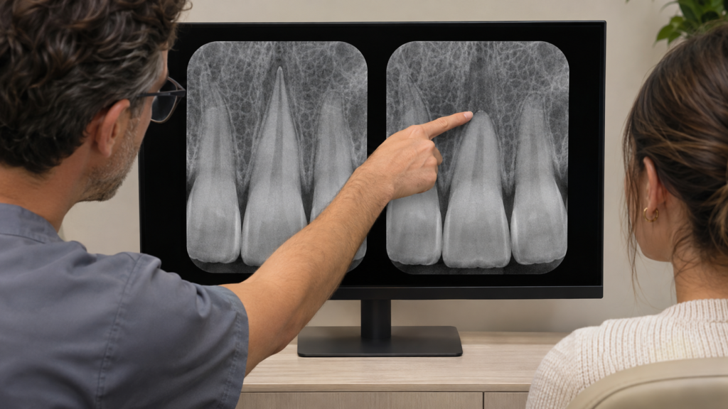
If your orthodontist says a tooth root looks shorter on an X-ray, ask to see the earlier image alongside it. The comparison helps show whether the root has changed and whether that change could affect your next adjustment.
Quick answer
Orthodontic root resorption is the loss of hard tissue from a tooth root during orthodontic tooth movement, typically at the tip. It usually causes no symptoms, so an orthodontist checks for it on dental X-rays when imaging is clinically indicated. A root that has shortened stays shorter, although the process usually settles once active force on the affected tooth stops. Your orthodontist may continue monitoring, change which teeth are moved, pause active movement, or end active treatment, depending on the findings.
What happens to the root in orthodontic root resorption?
The crown is the part of the tooth you see above the gum. The root sits below it, anchored in bone by a thin ligament. During orthodontic treatment, forces applied through an appliance allow teeth to move as the tissues around them remodel. In some teeth, cells also remove a small amount of hard tissue from the outside of the root. If that happens at the tip, the root can look shorter or more rounded on an X-ray. The technical term is external apical root resorption. “Apical” refers to the root tip.
Even when the root tip changes, the crown can look the same in the mirror. Small changes usually have little effect on long-term tooth health. Extensive shortening is uncommon and deserves a closer look because it can affect how well the tooth is supported, especially if the bone around it is also compromised. The British Orthodontic Society’s guidance on treatment risks describes this range and notes that individual susceptibility is difficult to predict.
Does root resorption from braces cause symptoms?
Usually, people cannot feel orthodontic root resorption. The European Society of Endodontology’s position statement on root resorption describes this type of external root resorption as typically symptom-free and notes that the tip can appear blunt on an X-ray. Feeling comfortable gives you little information about root length. A tooth that becomes noticeably loose or develops persistent tenderness deserves an examination because several dental issues can cause those changes.
If the term “external root resorption” appears in a report, ask where on the root the change is. External resorption describes several conditions. A finding near the gumline or a finding linked to infection may need a different assessment and a different kind of care from shortening at the root tip during tooth movement.
How do orthodontists monitor root shortening?
Orthodontists look at the root’s shape and length on available X-rays and compare earlier and later images when they can. The first image helps distinguish a root that started out short or rounded from a root that changed during care. At an orthodontic consultation, mention any previous finding of short roots or a significant dental injury, and tell the team if your dentist or a previous orthodontist may have relevant images.
A panoramic X-ray can give a broad view of the teeth. When it leaves a root tip hard to see, a targeted X-ray called a periapical radiograph may give the orthodontist a clearer view of the tooth and the surrounding bone. Image angle and clarity matter. A tip that appears shorter on a single image sometimes needs a better view or an earlier image for comparison.
There is no single repeat X-ray schedule for everyone. The clinical practice guideline on orthodontically induced root resorption recommends considering follow-up imaging in selected situations and taking a targeted view when existing images cannot adequately show root shape and size. The timing depends on what the orthodontist needs to check and whether the result could change the care plan.
Three questions to ask if your orthodontist spots a change:
Which tooth is affected, and can you show me the same tooth on my earlier X-ray?
How much root and supporting bone remain, and how confident are you in the comparison?
What would you need to see before moving this tooth further?
What does root resorption treatment involve?
For orthodontic root shortening, the goal is to limit further loss while considering what bite or alignment improvement is still worth pursuing. An orthodontist may keep monitoring a small finding, adjust the plan so a vulnerable tooth is no longer moved, or temporarily pause active tooth movement before reassessing. When significant shortening affects many teeth, ending active treatment early may be appropriate. If movement resumes, the orthodontist may recommend another X-ray when it can help determine whether the change is progressing.
The decision is specific to the tooth and the patient. For example, if a front tooth has already reached a useful position, moving it further for a small alignment improvement may offer little benefit compared with protecting the remaining root. An in-person examination and a review of the images are needed to weigh those options. If shortening is significant, your orthodontist can share the finding with your dentist so they can pay particular attention to the gums and bone supporting the affected teeth at future visits. These decisions draw on the clinical guideline above, whose recommendations for modifying treatment and providing follow-up care rely substantially on expert consensus.
Is root resorption reversible?
A root that has visibly shortened does not grow back to its original length. Small defects in the surface layer of the root, called cementum, can undergo some surface repair after active force is removed. That surface repair can help stabilize the area without restoring the lost length. An orthodontist may use follow-up records to see whether the root tip continues to change after the treatment plan is adjusted.
FAQs
One image can show root shape and length at a single point in time. An earlier image may show that the root already looked short, or a clearer targeted image may clarify an apparent difference. Your orthodontist will interpret the images alongside the examination.
A tooth with a shortened root can remain functional. The outlook depends in part on how much root remains, the condition of the gums and supporting bone, and whether the tooth is unusually mobile. These are reasons to keep regular dental follow-ups.
A root canal is not routinely used for shortening caused by orthodontic forces. If your dentist or orthodontist finds a separate problem involving the tissue inside the tooth, such as infection, that finding may need its own treatment.
Short roots are one factor in planning. An orthodontist will look at how the teeth are supported and how far they would need to move to achieve your goals. The findings may affect which movements are appropriate and how the roots are monitored.
Bring earlier and recent dental X-rays if they are available, or ask your previous provider to send them. An orthodontist can use those records and an in-person examination to assess the root’s condition and explain which movements may still be appropriate.